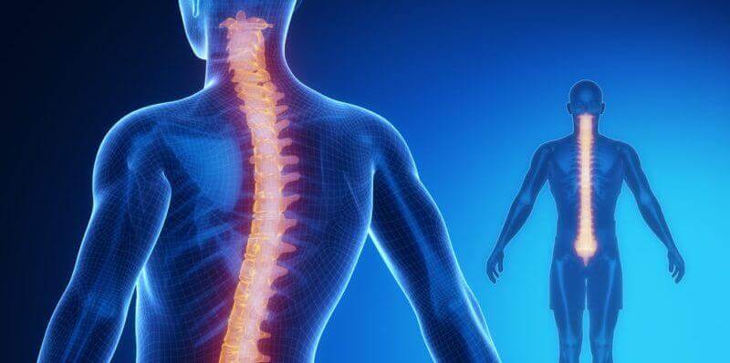
The spinal cord is part of the central nervous system (CNS), along with the brain, and extends from the occipital hole in the skull to about the first lumbar vertebra.
Along the spinal cord are connected 31 spinal nerves, consisting of a gray matter nucleus where the neural bodies are located, which in turn is surrounded by a white substance where the axons are located, curiously the distribution of gray and white matter in the spinal cord is the opposite of in the brain. On the other hand, as protection of the spinal cord, there are vertebrae, bra ligaments, meninges and cerebrospinal fluid.
- Spinal cord functions are varied.
- On the one hand it is responsible for receiving and processing (superficially) sensory information and.
- On the other hand.
- Sending motor information from the brain.
- Whose functions are essential and vitally important.
- An injury can cause serious effects such as motor paralysis or loss of sensation.
Gray matter, unlike the brain, is located inside the spinal cord, where neural bodies are located and where information is processed, is configured in different horns: ventral, dorsal, lateral and intermediate.
In addition, in this gray substance, there are several nuclei with different functions:
Gray matter in the spinal cord is the place to replace motor and sensory information, but it is also necessary to make a quick judgment about the information before it reaches its destination, the latter is necessary to activate the reflex mechanism in emergency situations, such as when receiving a very painful stimulus.
The white substance in the spinal cord is where the fibers (axons) that send information both up and down are located, their main function is to send information, like the black substance, is also divided into different parts, in this case columns:
In white matter there are different paths, ascending and descending, brochures or brochures bear the names of the two structures between which the information circulates, and each of the areas sends different information.
Thus, the white substance of the spinal cord is responsible for the transmission of motor and sensory information in a wide range of movements and sensations, communicating multiple areas.
The ascending pathways, as the name suggests, are responsible for sending information collected by the external senses (exteroceptive information) or internal (propioceptive) stimuli to the cerebral cortex, where deeper processing will be performed. Most upward pathways are elevated in the thalamus, with the exception of olfactory stimuli that go directly to the olfactory bulb.
They are ascending, centripetal, born from the periphery and providing information to the upper centers, some of the nerve fibers are used to connect different segments of the spinal cord, while others ascend from the spinal cord to the upper centers and connect the spinal cord to the brain. These pathways transmit information that may or may not reach consciousness.
In its simplest form, the pathway to consciousness consists of three neurons, but some related pathways use more or less. Many neurons in the ascending pathways branch out and others participate in reflex muscle activity.
These are the routes that carry the information of the somatoreceptors. There are two main routes:
Pyramidal pathways are the downward (motor) nerve pathways that pass through the pyramids. They are responsible for a fast, agile, fine and precise voluntary movement. There are three neurons involved in sending information to perform movements. They follow the following circuit:
The extrapyramidal pathway is responsible for involuntary movements, it comes from a subcortical structure that travels to the spinal cord. Regulates the execution of involuntary movements (walking, posture, muscle tone, vigilance and instinctive behavior). Unlike the pyramidal system, it does not begin in the cerebral cortex, but in several subcortical structures.
Another essential function of downstream motor pathways is to modulate the reflex circuits of the spinal cord. The adaptability of spinal reflexes may change depending on the behavioral context, because sometimes the gain (strength) or even the signal (extension vs bending) of a reflection must be modified so that the movement adapts to the circumstances, the descent lanes are responsible for controlling these variables.
There are movements that we perform unconsciously, before sensory information about the stimulus that causes movement reaches the brain, are reflex movements, such as removing the hand from a source of pain (for example, the flame of a lighter), or closing the eyes. when you hear a loud noise; we don’t control them.
The reflex is the simplest circuit of the nervous system, it begins at the level of the receptors, which are structures that transform the energy of the stimulus into an electrical change of the related peripheral nerves that lead the impulses to the center of integration, interneurone. The information passes to the efferent motor neurons, so that the effector (muscle) performs the reflex movement.
These movements occur through the reflex arc. The soma neuron is located in the node of the posterior root, passes through the dorsal, where it communicates with the interneurone, which is the association neuron that integrates information and passes to the motoneurone in the ventral pole to exit through the ventral root and direct the nerve impulse to the muscle for contraction.