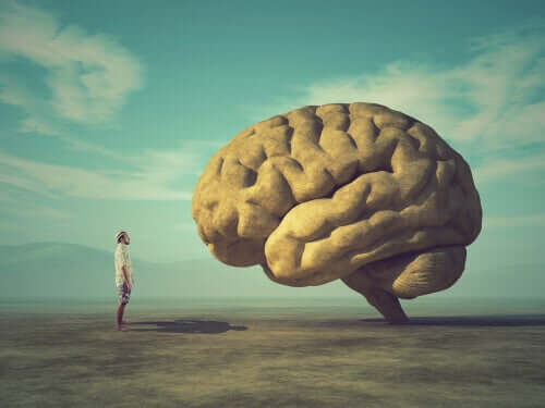
Throughout human history we wanted to know the brain and each of its parts, we also wanted to know what the function of each of its regions is and even know its most unknown place, today we want to continue looking for answers. , and the question we ask ourselves in this article is: why is the brain wrinkled?
Judging by the zoological scale, the relationship between neurons and learning capacity is linear; the larger the surface area of the brain, i. e. the larger the cerebral cortex, the greater the learning. Humans have the maximum surface area and their brains are wrinkled by brain convolutions. In this way, it is possible to have a large area in a small space.
- Animals without girth are called the lysencephalon and are unlikely to learn.
- While animals called leashes.
- With girth.
- Can learn more easily.
- The human being is the being most observed in time; its brain is more wrinkled than all other species.
- Followed by anthropoid monkeys.
“The human brain is composed almost exclusively of the cerebral cortex. A chimpanzee’s brain, for example, also has a bark, but in a much smaller proportion. The bark allows us to think, remember, imagine, are we human?” beings, essentially, by virtue of our cerebral cortex?. – Edoardo Boncinelli-
In May 2009, scientific american magazine published an article by Katherine S. Pollard, a biostatistician at the University of California, who developed software to compare segments of greater difference between humans and chimpanzees. The sequence that showed the biggest difference in this analysis has 118 nucleotides, which was called HAR1,?accelerated human region? (accelerated human region).
It appears that HAR1 has activity in the human brain and other vertebrates, this region has evolved very slowly in non-human vertebrates, since there are only two changes between nucleotides in chicken and chimpanzee sequences, while the number of differences between humans is 18.
The HAR1 sequence has been shown to be a modulator of gene expression in cultured cells, and it has been found to be active in a type of neuron involved in the development of the cerebral cortex, so when cells where HAR1 is activated are damaged, the brain develops abnormally and the cerebral cortex does not have its characteristic appearance (with many furrows and lobes).
An anatomical feature of intelligence is not only the weight of the brain, but the fact that it is wrinkled, that is, the amount of grooves and lobes it has.
“The spirit that opens up to a new idea will never return to its original size. “- Albert Einstein-
One of the most striking features of our brain is the incredible size of the cerebral cortex and its folds, visible as bumps and grooves on its outer surface.
As we mentioned, most animals with large brains have a folded cortex, while most animals with small brains have a smooth, crease-free bark.
The cerebral cortex is a laminar tissue where neurons are at the top. The lower or inner part contains most of the connection between neurons and areas of the brain.
In large brains, this layer of neural tissue covering the outside of the brain is disproportionately larger than the deep brain structures it covers and, instead of adopting a balloon shape, bends, minimizing the total volume of the brain and skull.
Victor Borrel and his team have been studying this field for several years and have shown that basal radial cilia (bRG), regulated by intrinsic and extrinsic factors, play a key role in tangential expansion of the cerebral cortex.
Thus, bRG is an essential, but not sufficient, requirement to generate a curved cortex, as it provides new radial processes through which neurons migrate radially, disperse tangentially and expand the cerebral cortex.
“The brain of the human baby, unlike any other animal, triples in size in its first year. “Morton Hunt.
Cortical folding occurs during embryonic development. The wrinkled form of the brain develops around the twentieth week of gestation and is completed when the child is a year and a half old.
This is important for optimizing functional organization and brain connections, and allows you to adjust a large cortex to a cranial volume limited by its size.
Some of the most common pathologies are polymicrogiria (formation of several small convolutions) and periventtricular nodular heterotopia, in the latter, neurons accumulate ectopically near the telecellic ventricles, forming nodules that act as epileptic focus.